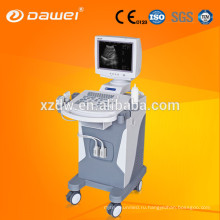


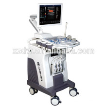
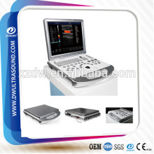
DW-C900 4d color doppler ultrasound machine & 4d color doppler usg
Базовая информация
Модель: DW-C900
Описание продукта
Product Description DW-C900 4d color doppler ultrasound machine & 4d color doppler usg

Mode of display Black-and-white image: B, 2B, 4B, right/left B|M, B|D, right/left PW(D), top/down PW(D), M, B mode local amplification, linear probe trapezoidal imaging, 3D reconstruction/real-time 3D imaging
Blood flow images: spectrum: B/BC, BC/B, B/D, B/C/D CFM: B|C|D, B|C|M, B|C double real-time, PW(D)(D), CFM, CPA, CW
Color Doppler:
Mode of display: energy, velocity, variance (in support of velocity variance), frequency, gain
Display control: the size and position of the sampling box are adjustable, the emitting angle of the linear probe is adjustable.
Spectral Doppler:
Sampling volume and position are adjustable
PW(D) tracing supports maximum and average velocity. The threshold of the maximum velocity is adjustable. Scanning scope: above base-line, below base-line and entire scanning.
PW(D) sound can be turned on or off, and can be regulated in volume.
Velocity measuring: in support of maximum and minimum velocity
Parameters for control of blood flow color images: Doppler frequency, position and size of sampling box, base line, color gain, deflection angle, wall filter, accumulative number of times, etc.
Color depth of display Continuously adjustable
Focusing Electronic focusing + sound lens focusing
Emission involves single focus, double focuses, triple focuses, and quadruple focuses Continuously dynamic focusing in receiving
Formation of digital beam Continuously dynamic focusing
Dynamic aperture diameter
Weighted sum of reception delay
In support of semi-moment scanning, and ±10° linear reception deflection angle
Multi-beam processing
Signal processing/Doppler sound Dynamic wave filtering
Quadrature demodulation
Total gain control
Composite gain technology: 8-section TGC and D-AGC.
Low-pass filter
Secondary sampling
Acoustic output adjustment
Adjustable Doppler stereophonic acoustic output
Image processing Dynamic scope variation and logarithmic compaction
Time filtering
Space filtering
Frame average
Fringe enhancement
Gray scale variation
B|M- or M-mode scanning speed
Linear density control
Scanning angle/width control
Image optimization
Upward and downward Image flipping
Multi-angle image rotation
Options for color gray-scale bar inversion
Combined processing of tissue and flow images
Resolution of black-and-white images: 1024×768×8 bits (256 gray-scale)
Resolution of color images: 8 bit×8 bit×8 bit color coding
Selection of specific images Optimized items are available to choose from
Basic measuring and calculating functions 2D normal measurement: distances, angle, perimeter, area (ellipse and locus methods), volume, volume – 3-line method (length, width, height), histogram, cross-section chart
Basic M-Mode measurement: time, distance, heart rate, valve rate
Obstetric measurement With 4 varieties of obstetric databases for reckoning gestation age
3 non-editable versions: Asian, European and American, plus 1 editable version: user-customized.
The gestation age can be reckoned by the information in each version on gestational sac diameter, crown-rump length, biparietal diameter, head circumference, abdominal circumference, femur length, humerus length, abdominal transversal diameter, length of vertebrae, occipital-frontal diameter, cerebellum diameter, fibula length, radius length, Cisterna Magna, inner orbital diameter, output orbital diameter, amniotic fluid index, fetal heart rate, nuchal transparency, distance and area, etc.
Obstetric report
Measurement and calculation of amniotic fluid index (AFI)
Ratio calculation (BPD/OFD, FL/AC, FL/BPD and HC/AC)
Fetus weight estimation
Reckoning the gestation age and EDD by LMP and BBT
Fetal biophysical score
Fetal growth curve
Andrology measurement and calculation Measurement and calculation of prostate and testis
Calculation of predicted prostatic specific antigen (PPSA) and Prostatic Specific Antigen density (PSAD).
Gynaecology measurement Measurement and calculation of the uterus, left ovarium, right ovarium, left folliculus, right folliculus, etc.
Urology measurement and calculation Detection and calculation of the left kidney, right kidney, bladder, residual urinal vol, etc.
Vessel measurement and calculation Measurement and calculation of area stenosis rate and vascular stenosis rate.
Multi-fetus detection and measurement Detection of multiple fetuses
Small parts detection and calculation Detection and calculation of thyroid, galactophore, masses, etc.
Orthopedic surgery measurement Detection of the left and right hip joint
Cardiac detection and calculation functions Cardiology measurement software package—contains features of analyzing and measuring aorta, mitral valve,( 54798395,left/right) ventricles, etc.
Area stenosis percentage (% area sten), tube diameter stenosis percentage (% Diam Sten)
Body surface area (BSA)
Body mark In terms of bodily function, an body mark database is divided into several parts: abdomen, andrology, cardiology, gynecology, musculoskeletal, obstetrics, pediatric, small parts, urology, etc.
Screen-displayed information Probe conditions, mode of display, depth, focus, dynamic range, body marks, probe position marker, acoustic output, patient conditions, name of medical agency, measured value, time and date, scale, scanning direction, gray-scale curve, current working frequency of the probe, frame frequency, B-Mode total gains, C-Mode total gains, Mode-D total gains, menu, annotations, gray-scale belt, puncture guide line, TI (thermal index), MI (mechanical index)
Storage Probe parameter storage
Image storage
Cine loop storage
Measurement results storage
Report storage
Cine loop Automatic cine-loop
Manual cine-loop
Cine-loop speed setting
Cine-loop search
Frame-by-frame forward/backward cine-loop
Digital ultrasonic working station (optional) General report
Obstetric report
Andrology report
Gynaecology report
Urology report
Blood vessel report
Small parts report
Cardiology report (ventricular report, mitral value report, aortic report, left ventricle report)
Search
Gray-scale 256 gray-scale
Language interface Chinese and English available
Function of the trackball Performing various operations based on the working state and menu
Controlling multiple electronic scale, locating the cursor, completing measurement
Input/output port VGA output port
DVI output port
Network port
USB port
Video port
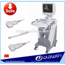
Introduction to the probe 3.5MHzR65 multi-frequency convex probe The probes have a frequency range between 2.0MHz-5.5MHz. In the B-Mode , they have a scanning angle of 60°. In the M-Mode, they can activate particular array element as fixed-frequency probes. They can be used in the combination of B-Mode and M-Mode.
Scope of application: examination and diagnosis of abdominal tissues, such as liver, gallbladder, spleen, pancreas, kidneys, gastro-intestinal, uterus, urinary bladder, prostate, etc.; assessment of growth and maturity of fetus in obstetric diagnosis.
6.5MHzR10 multi-frequency convex probe The probes have a frequency range between 5.0MHz-9.0MHz. In the B-Mode , they have a scanning angle of 140-158°. In the M-Mode, they can activate particular array element as fixed-frequency probes. They can be used in the combination of B-Mode and M-Mode.
Scope of application: vagina, uterus, rectum, rectum wall, prostata, etc.
Warning: Oil- and bacteria-free rubber sleeve must be added when using the probes. The probes should not be activated until going in the patient’s body.
7.5MHzL46 multi-frequency linear probe The probes have a frequency range between 6.0MHz-12.0MHz. In the B-Mode , they have a scanning width of 38-45. In the M-Mode, they can activate particular array element as fixed-frequency probes. They can be used in the combination of B-Mode and M-Mode.
Scope of application: newborns, musculoskeletal system, peripheral vessels, small parts (including breasts, thyroid, testis, etc.)
3.0MHzL19 multi-frequency phased-array probe The probes have a frequency range between 2.0MHz-5.5MHz. In the B-Mode , they have a scanning angle of 90°. In the M-Mode, they can activate particular array element as fixed-frequency probes. They can be used in the combination of B-Mode and M-Mode.
Scope of application: heart, abdominal tissues and parts
4.0MHzR40 multi-frequency convex volume probe The probes have a frequency range between 2.0MHz-5.5MHz. In the B-Mode , they have a scanning angle of 70°. In the M-Mode, they can activate particular array element as fixed-frequency probes. They can be used in the combination of B-Mode and M-Mode.
Scope of application: real-time 3D scan; examination and diagnosis of fetus, abdominal tissues, uterus, etc.; assessment of growth and maturity of fetus in obstetric diagnosis.
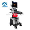
4d color doppler images:


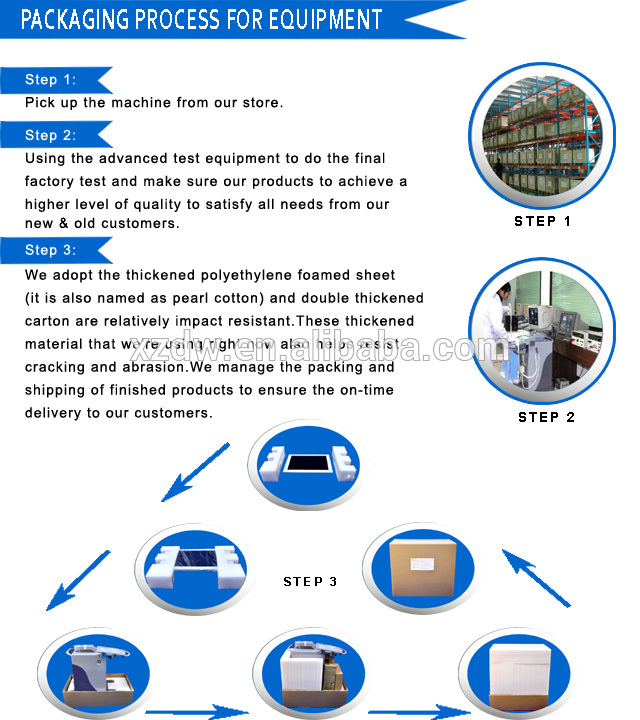
Packaging & Shipping 4d color doppler packing details:
 Delivery details:
Brand |
DAWEI |
Origin |
China |
MOQ |
1 set |
Delivery |
3(THREE) working days after receiving the payment |
Payment |
TT,Western Union,Paypal,Escrow, L/C,Credit Sale |
Package |
Packed double cartons,bound with four plastic belts externally |
Warranty |
TWO years warranty . After TWO years the cost of providing paid services to maintenance |
Validity |
7 working days |
Company Information


If you want to know more about our 4d color doppler, contact us now!!
Группа Продуктов : 4Д/3Д Цвет doppler машины ультразвука
Другие продукты


Горячие продукты
медицинский портативный цвет doppler машины ультразвукаКлиника мочевого пузыря сканер цен & портативный УЗИ аппаратвагонетка УЗИ 4D цена сканер цветной допплер ecografos с свободной руки 3D и 4D УЗИ ценаНовое Прибытие 19" плюс 10.4"реального времени СИД 4Д цвет doppler вагонетки высокого класса 3D и 4D УЗИ машиностроительные заводыцена машины ультразвука и ультразвуковой аппарат для беременностиДГ-350 низкой цене польностью цифровой блок развертки ультразвука вагонеткиДГ-C60plus цвет доплеровский ультразвуковой машины портативный 3D 4D для продажиVET6 блок развертки ультразвука ладони ДГ-360 высокое качество изображения портативный ультразвуковой аппарат ценаДАВЭЙ ДГ-500 самые дешевые в 2D Китай портативный ультразвуковой аппарат ценаДавэй ДГ-350 B вагонетки ультразвуковая диагностика машиныпортативный блок развертки ультразвука handheld машина доплер УЗИ ветеринарный портативный УЗИ свиньи для беременностиветеринарный УЗИ верблюд беременности ДГ-C80plus двойного экрана 4D doppler цвета цифров ультразвуковая машина ультразвуковая машина горячей продажу в ЕгиптеДГ-С80 4D УЗИ машина doppler цвета & экономичный уздг